43 microscope images with labels
DP Biology: Ultrastructure of cells quiz 1.2 - ThinkIB It means that the diameter of the resolved image will be 0.1µmm. It means that the distance from the objective lens to the slide should be 0.1µm. It means that the largest thing visible in the microscope is 0.1µm. It means that 0.1µm is the smallest distance which two objects can be seen as separate objects. Microscopes, Software and Imaging Solutions from Carl Zeiss USA Your Partner in cutting-edge microscopy. As a leading manufacturer of microscopes ZEISS offers inspiring solutions and services for your life sciences and materials research, teaching and clinical routine. Reliable ZEISS systems are used for manufacturing and assembly in high tech industries as well as exploration and processing of raw ...
Microbiology Virtual Lab I - Amrita Vishwa Vidyapeetham Fig:-Lophotrichous flagellum seen under light microscope . Flagella are spread fairly evenly over the whole surface of peritrichous bacteria . Fig:-Peritrichous flagellum seen under light microscope . When anticlockwise rotation is resumed, the cell tends to move in a new direction. This ability is important, since it allows bacteria to change ...

Microscope images with labels
The Whole-transcriptome Landscape of Diabetes-related Sarcopenia ... The decreased CSA and numbers of myotubes were observed in PA-treated C2C12 cells through fluorescently labeled MyHC by confocal microscopy (a) and light microscopy (b). Georeferencing Topo Sheets and Scanned Maps (QGIS3) - QGIS Tutorials The Georeferencer is divided into 2 sections. The top section where the image will be displayed and the bottom section where a table showing your GCPs will appear. Now we will open our JPG image. Go to File ‣ Open Raster. Browse to the downloaded image of the scanned map and click Open. MeCP2 inhibits ischemic neuronal injury by enhancing methylation of the ... The sections were observed under a light microscope, and the apoptosis index was subsequently calculated. Immunofluorescence assay Cell counting Kit-8 (CCK-8) assay
Microscope images with labels. Critics say 'monkeypox' is a racist name. But it's not going away ... Nearly seven weeks after the World Health Organization said it will change the name of the monkeypox disease, agreeing with scientists who called it "discriminatory and stigmatizing," the controversial label doesn't seem to be going anywhere.. Critics say the name "monkeypox" plays into racist stereotypes about Black people, Africa and LGBTQ people — and, they note, it falsely suggests ... Dark-field Microscopy: Principle and Uses - Microbe Online Principle. Dark-field microscopy uses a light microscope with an extra opaque disc underneath the condenser lens, or a special condenser having a central blacked-out area, due to which the light coming from the source cannot directly enter into the objective. The path of the light is directed in such a way that it can pass through the outer ... Critics say 'monkeypox' is a racist name. But it's not going away ... This 2003 electron microscope image made available by the Centers for Disease Control and Prevention shows mature, oval-shaped monkeypox virions, left, and spherical immature virions, right ... Microscopio Leica - leica compound microscope labeled micropedia ... Microscopio Leica - 17 images - microscopio leica dm500 bio optic s r l, microscopio vertical de materiales leica dm4 m aspelab, leica microscopes kdb healthcare, carl zeiss axiostar binocular microscope a personal review,
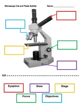
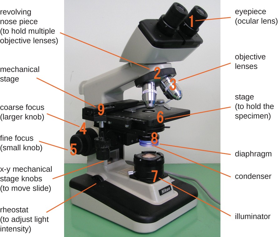
Print Resolution: The Essential Guide to Image Resolution for Printing So if you want to know how large a 24 MP full-frame camera can print at 300 PPI, you'll divide 6000 pixels by 300 PPI and 4000 pixels by 300 PPI, for 13.3 x 20 images. So, a 24 MP camera, using 300 PPI images, can print 13.3 x 20 images at maximum. And the only way to increase your print size is to drop the PPI, which will in turn hurt print ... ECLIPSE Ti2 Series | Inverted Microscopes | Products | Nikon ... The ECLIPSE Ti2 inverted microscope delivers an unparalleled 25mm field of view (FOV) that revolutionizes the way you see. With this incredible FOV, the Ti2 maximizes the sensor area of large-format CMOS cameras without making compromises, and significantly improves data throughput. The Ti2's exceptionally stable, drift-free platform is ... Light Microscope (Theory) - Amrita Vishwa Vidyapeetham The modern compound microscope consists of two lens system, the objective and the ocular or eye piece. The first magnified image obtained with objective lens, is again magnified by the eye piece to give a virtual inverted image. The total magnification the product of the magnifications of two lens systems. Parts of a Microscope protozoan | Definition, Parasites, Diseases, Characteristics, Size ... protozoan, organism, usually single-celled and heterotrophic (using organic carbon as a source of energy), belonging to any of the major lineages of protists and, like most protists, typically microscopic. All protozoans are eukaryotes and therefore possess a "true," or membrane-bound, nucleus. They also are nonfilamentous (in contrast to organisms such as molds, a group of fungi, which ...
DENSsolutions - In situ Microscopy Innovative Solutions Aug 1, 2022. In a collaborative effort between the Dalian Institute of Chemical Physics (DICP) and DENSsolutions, a team of experts developed a data synchronization method to account for time delays in operando gas and heating TEM. Specifically, they systematically explored the relationship between delayed time and critical reaction conditions. Kidney histology: Nephron, loop of Henle, functions | Kenhub Kidney histology. The kidneys are paired retroperitoneal organs of the urinary system. Their function is to filter blood and produce urine. Each kidney consists of a cortex, medulla and calyces. The nephron is the main functional unit of the kidney, in charge of removing metabolic waste and excess water from the blood. Kidney Under Microscope Labeled - simple cuboidal epithelium ... Kidney Under Microscope Labeled - 17 images - histology microscope prepared slides, simple cuboidal epithelium youtube, primary hyperparathyroidism and minimally invasive parathyroid surgery, slide 67 testis male reproductive system youtube, N-SIM S | Super-Resolution Microscopes - Nikon Instruments Inc. The N-SIM S achieves incredible acquisition speeds (up to 15 fps*), enabling super-resolution time-lapse imaging of live cells and intracellular dynamics. * 2D-SIM mode, 512 x 512 pixels, 2 msec exposure time. Endosomes of a COS7 cell labeled with YFP. Rapid movement of endosomes is captured at high resolution.
Labeling a Compound Microscope Quiz
3D Laser Scanning Microscope, VK-X200 - KEYENCE Inline 3D Measurement: Connector 00:55. Inline 3D Measurement: BGA Ball 00:55. World's Smartest Fiber Sensor 1:34. 3D Laser Scanning Microscope 2:36. SR-2000 Series 0:53. WI-5000 Series 00:46. VR-3000 Series 01:15. Autofocus 1D and 2D Code Reader SR-1000 1:46. Version Upgrade 1:29.
32 Picture Of Microscope With Label - Labels For You
how to draw what you see in a microscope - Chet Gunther Up to 24 cash back Equipment You will need. Observe an onion cell under the microscope. Make sure that this line will overlap with the oblongs. Once you focused your image take your paper pencil and mathematical compass and draw what you see on the microscope Once you have finished drawing your image add labels to your drawing and a title.

Microscope Clip Art at Clker.com - vector clip art online, royalty free & public domain
Sportswashing Explained: What LIV Golf Means to the Saudi Government AP Photo/Seth Wenig. LIV Golf is an upstart league backed by the Saudi government meant to compete with the PGA Tour. Critics have accused LIV Golf as an example of "sportswashing," or using the love of sports to cleanse the image of an authoritarian regime, like Saudi Arabia. Whether or not sportswashing works depends on who you ask, and how ...
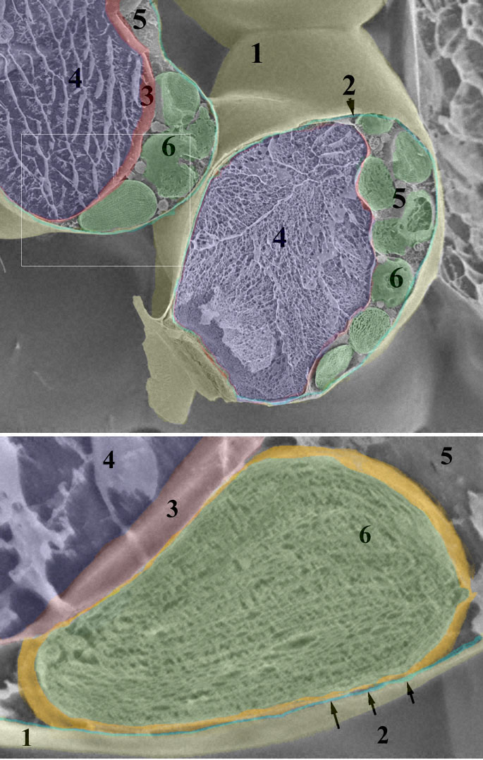
Leaf chloroplast
Opinion: COVID rises again, and now we have monkeypox AP file photo / This 2003 electron microscope image made available by the Centers for Disease Control and Prevention shows mature, oval-shaped monkeypox virions, left, and spherical immature ...

Labels Of The Microscope
13 Best Image Annotation Tools of 2022 [Reviewed] - V7Labs Hive Data is a fully managed data annotation solution to source and label training data for AI / ML Models. Hive Data supports image, video, text, 3D Point Cloud annotation and data sourcing. Apart from basic annotation types, Hive Data offers multi-frame object tracking, contours, and 3D panoptic segmentation.

MICROBIOLOGY SLIDE SPECIMENS
Is 'monkeypox' racist? CT official calls for a name change FILE - This 2003 electron microscope image made available by the U.S. Centers for Disease Control and Prevention shows mature, oval-shaped monkeypox virions, left, and spherical immature virions, right, obtained from a sample of human skin associated with the 2003 prairie dog outbreak.

Microscope labeling
Perseid meteor shower 2022: When, where & how to see it Perseid meteor shower: Quick facts. — When: July 14 to August 24. — Peak: Aug. 11-12. — Comet of origin: 109P/Swift-Tuttle. — Zenithal Hourly Rate (ZHR): 100. (The number of meteors a ...

Best Top Desktop Wallpapers: Electron microscope images
Mr. Jones's Science Class Earth, Moon, & Sun System (PPT.) Seasons Interactive (Online Activity) Moon Phases - Introductory Activity. Modeling the Phases of the Moon. Problems in Space (Online Activity) Lunar & Solar Eclipses - Webquest.
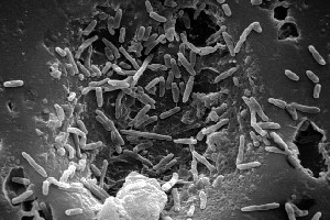
Mycobacterium leprae* - microbewiki
Optics News | Photonics News Researchers Develop Sprayless 3D Computer-Aided Verification Tool for Plastic. A recent study in Polymers suggests a novel, environmentally friendly three-dimensional technology for optical ...
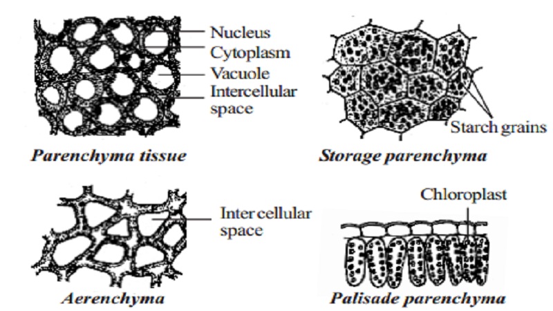
Permanent tissue: characteristics, types and functions - Online Biology Notes
Midsagittal section of the brain: anatomy - Kenhub The midsagittal section of the brain shows the three major parts of the brain, which are the cerebrum, cerebellum, and brainstem.The cerebrum (prosencephalon or forebrain) comprises the telencephalon (cerebral hemispheres) and the diencephalon.They are each also divided into subparts or regions for simplified localization of structures, for example, the brainstem is composed of the midbrain ...

31 Label The Indicated Parts Of The Microscope - Labels For Your Ideas
Biden Press Secretary Not Ready For Prime Time Jen Psaki, regardless of how you feel about Biden, did her job well. She spun decently and lied, lied, lied. Karine Jean-Pierre lacks that press secretary professionalism.
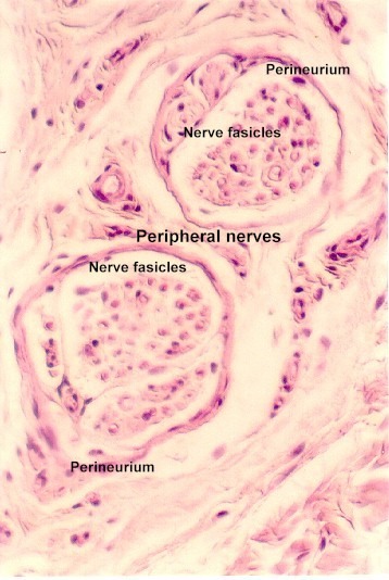
Peripheral Nerve Histology | Microanatomy Web Atlas | Gwen V. Childs, Ph.D.
MeCP2 inhibits ischemic neuronal injury by enhancing methylation of the ... The sections were observed under a light microscope, and the apoptosis index was subsequently calculated. Immunofluorescence assay Cell counting Kit-8 (CCK-8) assay
Tissues Flashcards | Easy Notecards
Georeferencing Topo Sheets and Scanned Maps (QGIS3) - QGIS Tutorials The Georeferencer is divided into 2 sections. The top section where the image will be displayed and the bottom section where a table showing your GCPs will appear. Now we will open our JPG image. Go to File ‣ Open Raster. Browse to the downloaded image of the scanned map and click Open.

32 Label Of Compound Microscope - Label Design Ideas 2020
The Whole-transcriptome Landscape of Diabetes-related Sarcopenia ... The decreased CSA and numbers of myotubes were observed in PA-treated C2C12 cells through fluorescently labeled MyHC by confocal microscopy (a) and light microscopy (b).

Microscope labeling
Microscope Introduction – “e” Lab - Biology LibreTexts
23 Label And Color The Parts Of Both Microscopes - Labels 2021
Post a Comment for "43 microscope images with labels"