45 fluorescent labels and light microscopy
Fluorescence Microscopy vs. Light Microscopy - New York Microscope Company Light microscopes use light in the 400-700nm range - the range through which light is visible to the human eye - but fluorescence microscopy uses much higher intensity light. Because traditional light microscopy uses visible light, the resolution is more limited. Fluorescence microscopy, on the other hand, uses light produced by the ... Fluorescent Lighting in Winnipeg, Manitoba - Kijiji™ Find "Fluorescent Lighting" in Winnipeg, Manitoba - Visit Kijiji™ Classifieds to find new & used items for sale. Explore Jobs, Services, Pets & more.
Learn about fluorescent labels and light microscopy | Echemi Provide different insights into fluorescent labels and light microscopy on echemi.com. We offer a huge of fluorescent labels and light microscopy news and articles here. ... Fluorescent Brightener. Plastic Rubber Chemicals. Polymer. Precious Metal Catalysts. Zeolite. Flame Retardants. Petrochemical. Natural Products. Lignans. Xanthones ...

Fluorescent labels and light microscopy
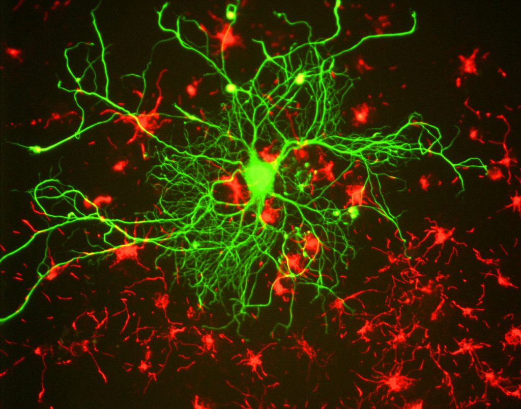
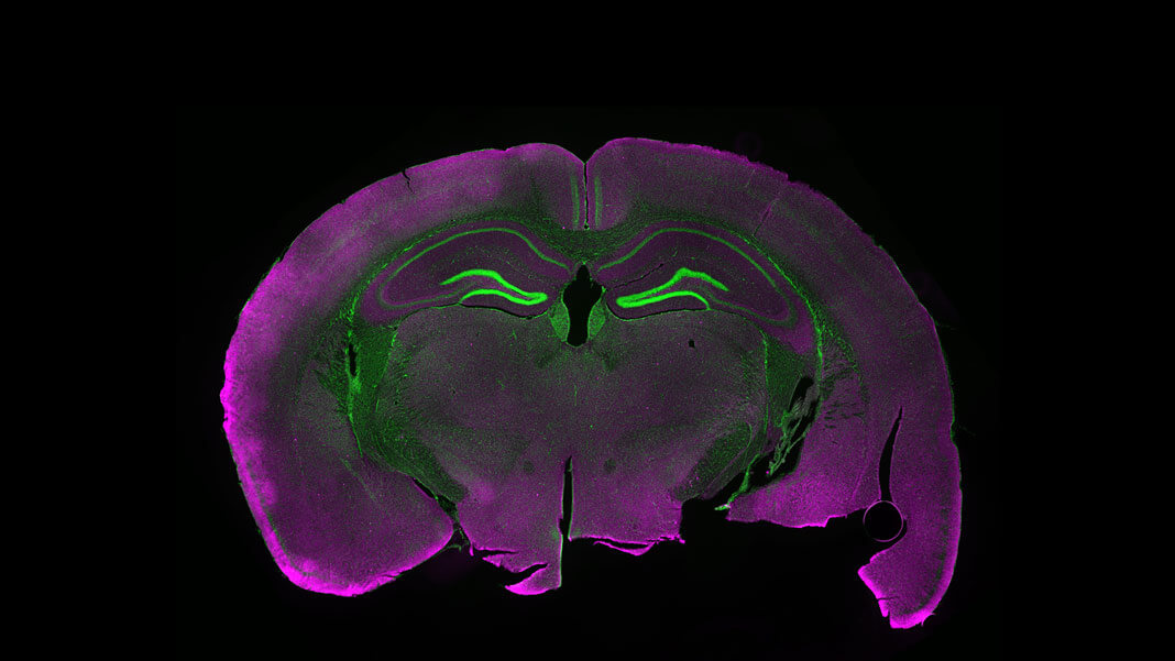
Fluorescence Microscopy vs. Light Microscopy - News-Medical.net This means that fluorescent microscopy uses reflected rather than transmitted light. For example, a commonly used label is green fluorescent protein (GFP), which is excited with blue light and... Fluorescent Microscopy Fluorescent microscopy is often used to image specific features of small specimens such as microbes. It is also used to visually enhance 3-D features at small scales. This can be accomplished by attaching fluorescent tags to anti-bodies that in turn attach to targeted features, or by staining in a less specific manner. Fluorescence Microscopy - Explanation and Labelled Images Fluorescence microscopy uses a high-intensity light source that excites a fluorescent molecule called a fluorophore in the sample observed. The samples are labeled with fluorophore where they absorb the high-intensity light from the source and emit a lower energy light of longer wavelength.
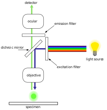
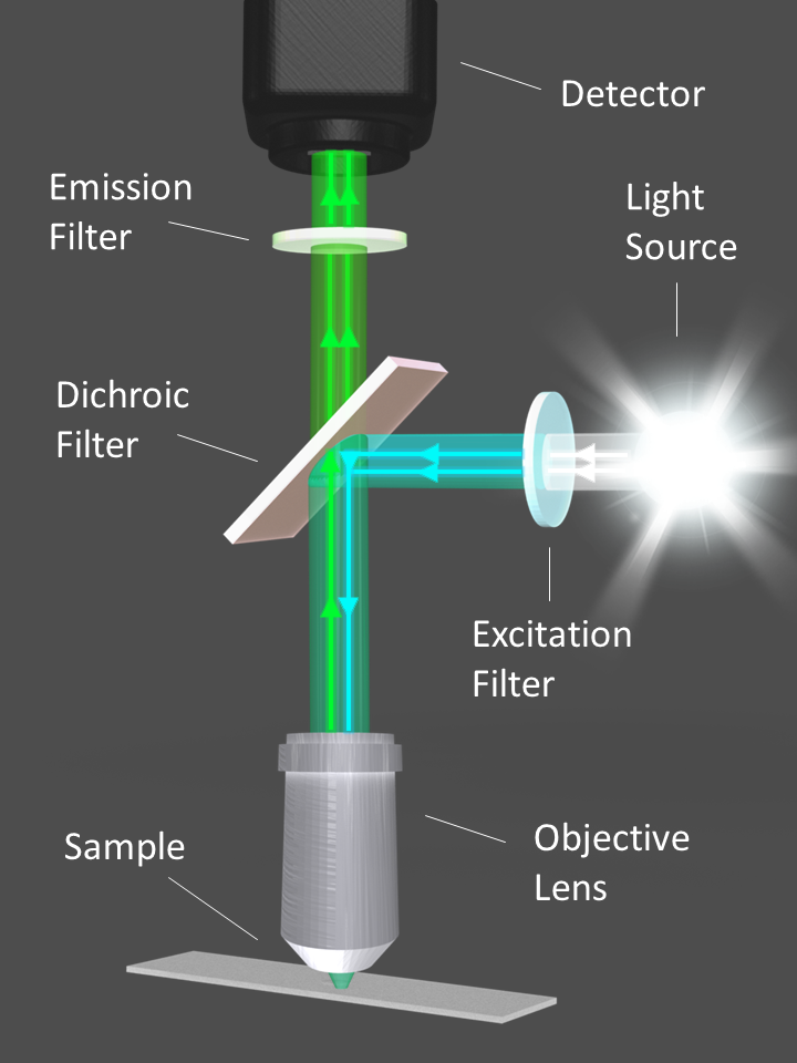
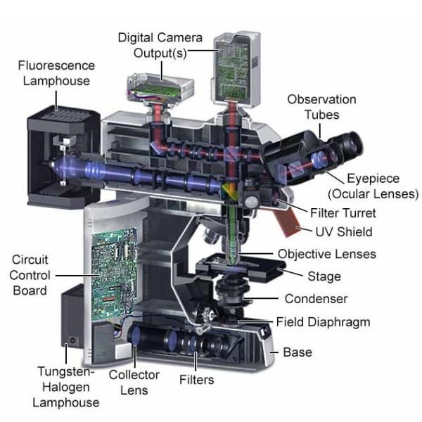
Fluorescent labels and light microscopy. Fluorescence microscope - Wikipedia The majority of fluorescence microscopes, especially those used in the life sciences, are of the epifluorescence design shown in the diagram.Light of the excitation wavelength illuminates the specimen through the objective lens. The fluorescence emitted by the specimen is focused to the detector by the same objective that is used for the excitation which for greater resolution will need ... Fluorescent Lights in Manitoba - Kijiji™ fluorescent light; fluorescent; light fixture; free; shop lights; tube light; led bulbs; vanity light; garage lights; Post an Ad. Manitoba > Results for "fluorescent lights" Results for "fluorescent lights" in All Categories in Manitoba - Page 2 Showing 41 - 80 of 101 results. Notify me when new ads are posted. Fluorescence Imaging - Teledyne Photometrics Fluorescent molecules (known as fluorophores) are used to label samples, and fluorophores are available that emit light in virtually any color. In a fluorescent microscope, a sample is labeled with a fluorophore, and then a bright light ( excitation light) is used to illuminate the sample, which gives off fluorescence ( emission light ). Label-free prediction of three-dimensional fluorescence images from ... We present a label-free method for predicting three-dimensional fluorescence directly from transmitted-light images and demonstrate that it can be used to generate multi-structure, integrated...
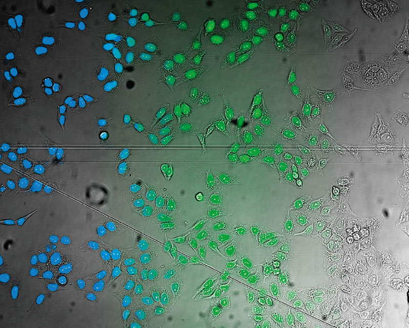
Label-free prediction of three-dimensional fluorescence images from ... Although fluorescence microscopy can resolve subcellular structure in living cells, it is expensive, is slow, and can damage cells. We present a label-free method for predicting three-dimensional fluorescence directly from transmitted-light images and demonstrate that it can be used to generate multi-structure, integrated images. Fluorescent Microscopy - ¿What is HPLC? In the 1930s the first fluorescent labels or probes for marking non-fluorescent objects appeared. In the 1950s Albert Koons and Nathan Kaplan developed a method for detecting antigens in tissues using antibodies labeled with fluorescent dyes. ... The main elements of a fluorescence microscope: Light source (mercury, xenon lamps, LED, laser); Fluorescent Label - an overview | ScienceDirect Topics Fluorescence microscopy is a very common tool. Usually, fluorescent labels are used to brighten up the object of interest. However, the same strategy is not applicable for graphitic materials, such as graphite, graphene, GO or r-GO as they are strong quenchers of dye molecules [45].Therefore, we developed a reverse strategy, FQM, where graphene-based sheets appear dark against a bright ... Fluorescent tag - Wikipedia S. cerevisiae septins revealed with fluorescent microscopy utilizing fluorescent labeling In molecular biology and biotechnology, a fluorescent tag, also known as a fluorescent label or fluorescent probe, is a molecule that is attached chemically to aid in the detection of a biomolecule such as a protein, antibody, or amino acid.
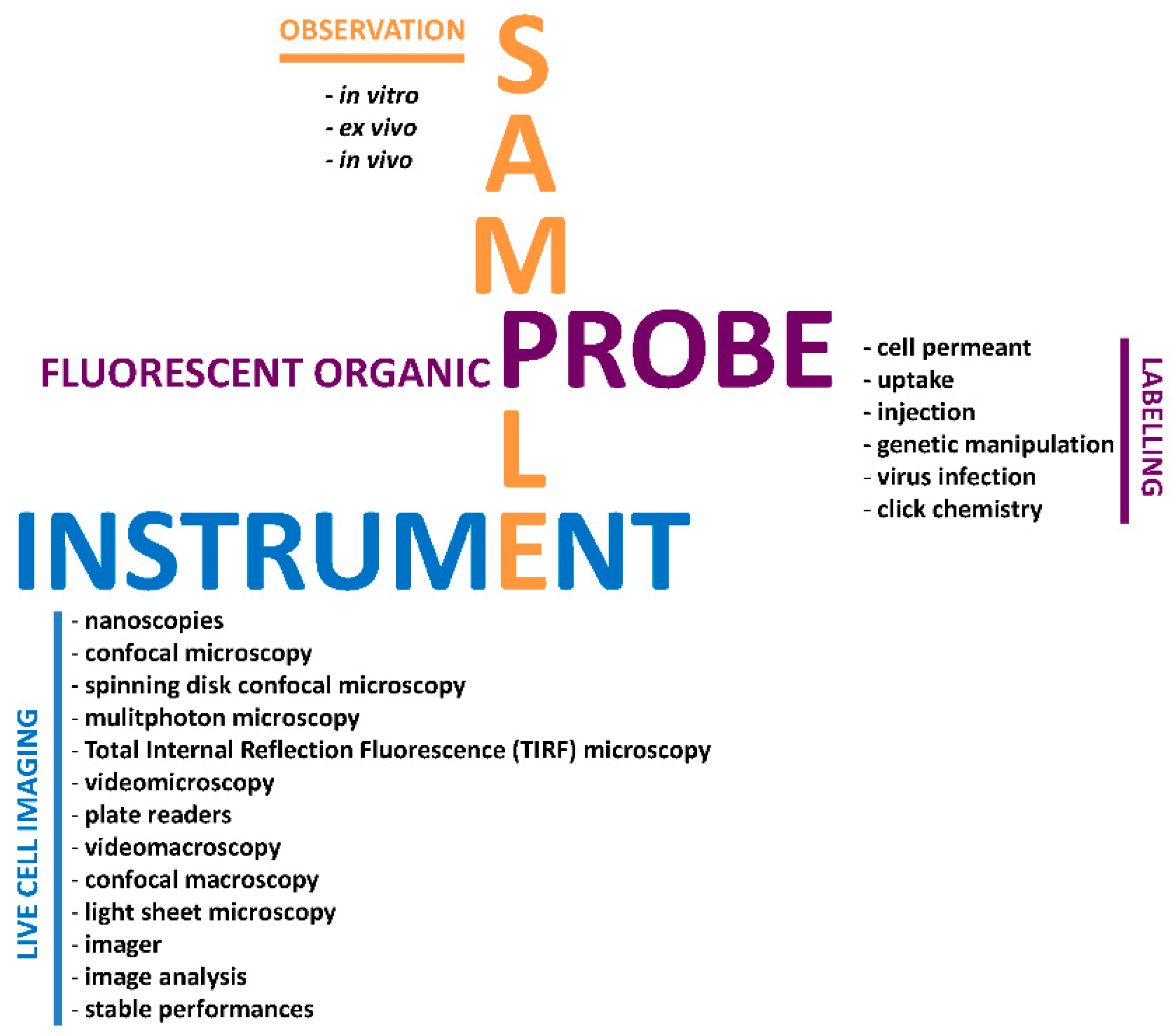
Fluorescence Microscope: Principle, Types, Applications Fluorescence microscopy is a light microscope that works on the principle of fluorescence. A substance is said to be fluorescent when it absorbs the energy of invisible shorter wavelength radiation (such as UV light) and emits longer wavelength radiation of visible light (such as green or red light). This phenomenon, also called fluorescence ... Different Ways to Add Fluorescent Labels - Thermo Fisher Scientific Learn about what characteristics of fluorescent dyes are important and see how they can be used in functional and structural cell analysis studies. Different Ways to Add Fluorescent Labels | Thermo Fisher Scientific - KR Why would scientists use fluorescent labels or dyes when using a light ... What is the advantage of fluorescence microscopy over light microscopy? Advantages of Fluorescence Microscope Fluorescence microscopy is the most popular method for studying the dynamic behavior exhibited in live-cell imaging. This stems from its ability to isolate individual proteins with a high degree of specificity amidst non-fluorescing ... WORKSHOP: "MICROSCOPY, OPTICS and IMAGING" | Bioscience Association ... We are glad to announce our next workshop on "Microscopy, Optics and Imaging", which will take place in Winnipeg (Manitoba) from November 16-20, 2015. TOPICS COVERED: - Basic Microscopy, Live Cell Imaging, Digital Imaging and Documentation, Automated Sample Analysis - Fluorescent Dyes, Probes, Instruments, Platforms and Applications
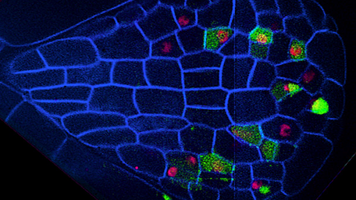
Labeling the ER for Light and Fluorescence Microscopy Here we describe methods of labeling the native ER using fluorescent proteins and lipid dyes as well as methods for immunolabeling on plant tissue. Labeling the ER for Light and Fluorescence Microscopy Methods Mol Biol. 2018;1691:1-14. doi: 10.1007/978-1-4939-7389-7_1. ...
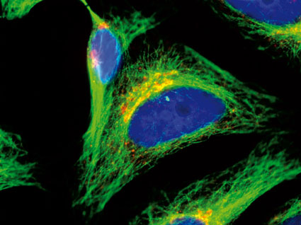
Fluorescence Microscopy - Explanation and Labelled Images Fluorescence microscopy uses a high-intensity light source that excites a fluorescent molecule called a fluorophore in the sample observed. The samples are labeled with fluorophore where they absorb the high-intensity light from the source and emit a lower energy light of longer wavelength.
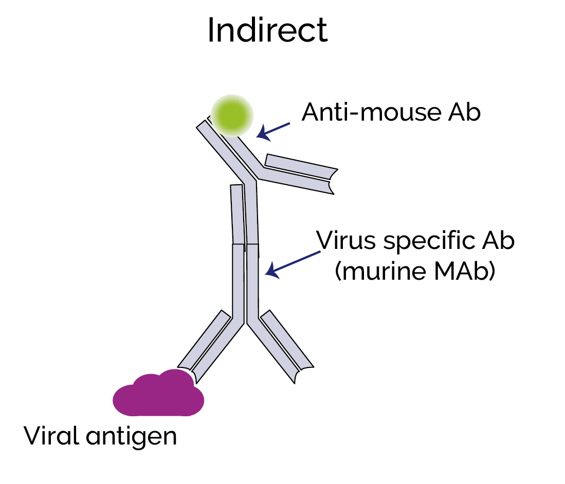
Fluorescent Microscopy Fluorescent microscopy is often used to image specific features of small specimens such as microbes. It is also used to visually enhance 3-D features at small scales. This can be accomplished by attaching fluorescent tags to anti-bodies that in turn attach to targeted features, or by staining in a less specific manner.
Fluorescence Microscopy vs. Light Microscopy - News-Medical.net This means that fluorescent microscopy uses reflected rather than transmitted light. For example, a commonly used label is green fluorescent protein (GFP), which is excited with blue light and...










Post a Comment for "45 fluorescent labels and light microscopy"