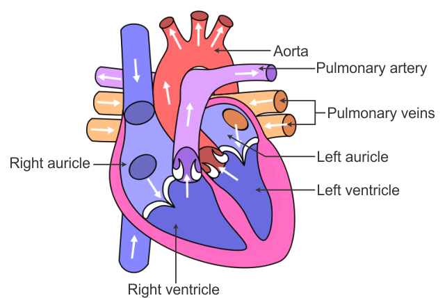
42 the human heart and its labels
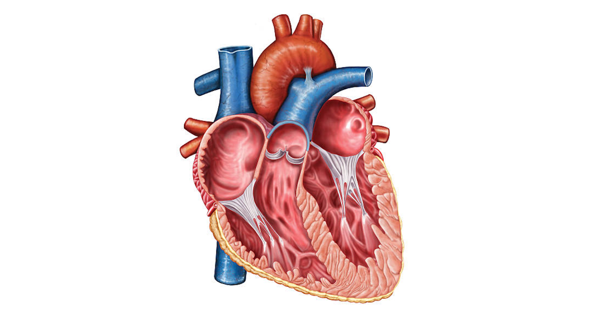
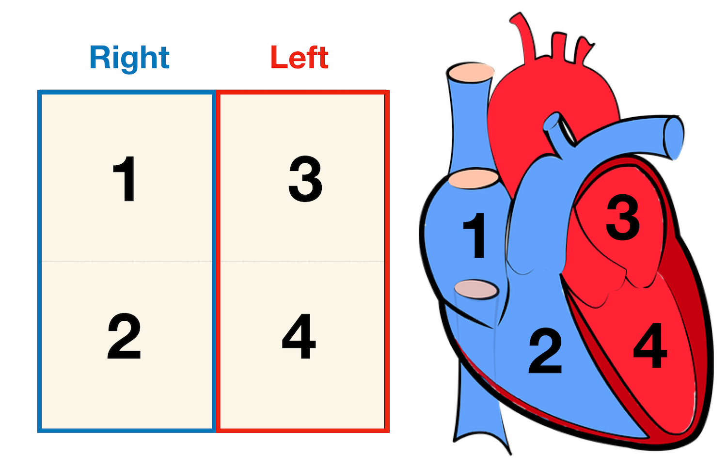
Heart - Wikipedia The human heart is situated in the mediastinum, at the level of thoracic vertebrae T5-T8.A double-membraned sac called the pericardium surrounds the heart and attaches to the mediastinum. The back surface of the heart lies near the vertebral column, and the front surface known as the sternocostal surface sits behind the sternum and rib cartilages. The upper part of the heart is the attachment ... Human Heart - Diagram and Anatomy of the Heart - Innerbody The heart contains 4 chambers: the right atrium, left atrium, right ventricle, and left ventricle. The atria are smaller than the ventricles and have thinner, less muscular walls than the ventricles. The atria act as receiving chambers for blood, so they are connected to the veins that carry blood to the heart.
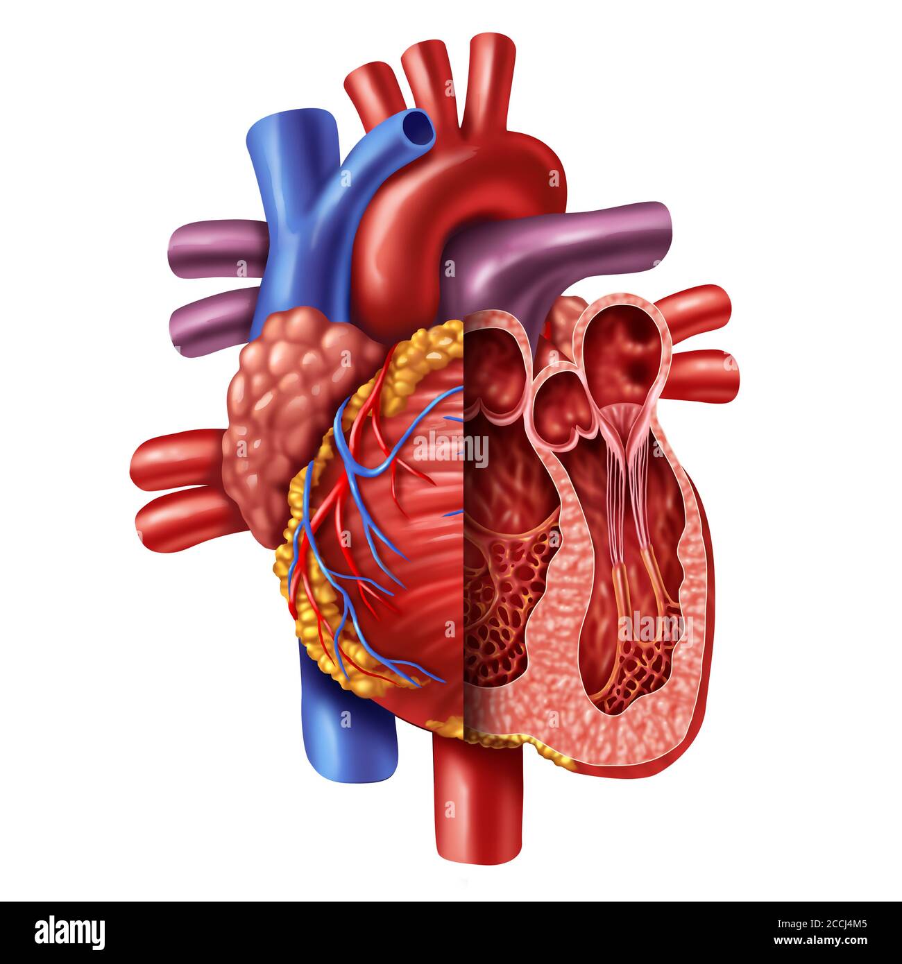
Anatomy of a Human Heart - U of M Health Located between the lungs in the middle of the chest, the heart pumps blood through the network of arteries and veins known as the cardiovascular system. It pushes blood to the body's organs, tissues and cells. Blood delivers oxygen and nutrients to every cell and removes the carbon dioxide and other waste products made by those cells.

The human heart and its labels
Prepare a drawing of the human heart and label its parts. | Quizlet Draw a diagram of the heart showing the three layers composing its wall and its four chambers. Label each. Show where the AV and semilunar valves are. Show and label all blood vessels entering and leaving the heart chambers. Human Genome Project Results Nov 12, 2018 · By using mRNA as a template, scientists use enzymatic reactions to convert its information back into cDNA and then clone it, creating a collection of cDNAs, or a cDNA library. These libraries are important to scientists because they consist of clones of all protein-encoding DNA, or all of the genes, in the human genome. Heart Pictures, Diagram & Anatomy | Body Maps - Healthline The average human heart weighs between 6 and 11 ounces. The muscle is strong enough to pump up to 2,000 gallons — as much as a fire department's tanker truck — of blood through one's body ...
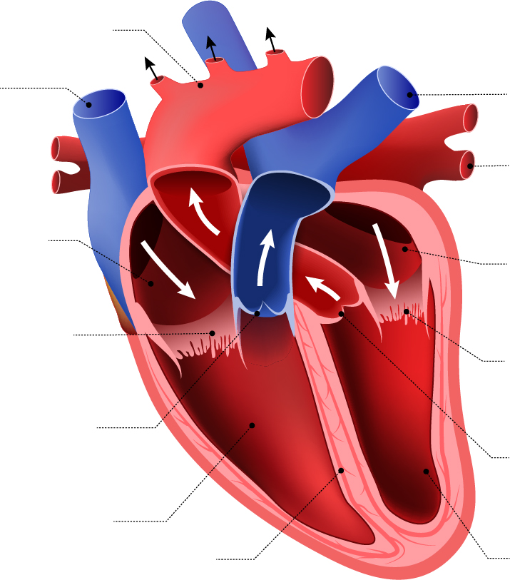
The human heart and its labels. File:Diagram of the human heart (no labels).svg - Wikimedia File:Diagram of the human heart (no labels).svg. From Wikimedia Commons, the free media repository. File. File history. File usage on Commons. Metadata. Size of this PNG preview of this SVG file: 498 × 599 pixels. Other resolutions: 199 × 240 pixels | 399 × 480 pixels | 639 × 768 pixels | 851 × 1,024 pixels | 1,703 × 2,048 pixels | 533 × ... 147 Heart Anatomy With Labels Premium High Res Photos - Getty Images Browse 147 heart anatomy with labels stock photos and images available, or start a new search to explore more stock photos and images. of 3. NEXT. Heart: Anatomy and Function - Cleveland Clinic Heart. Your heart is the main organ of your cardiovascular system, a network of blood vessels that pumps blood throughout your body. It also works with other body systems to control your heart rate and blood pressure. Your family history, personal health history and lifestyle all affect how well your heart works. Appointments 800.659.7822. Heart Diagram – 15+ Free Printable Word, Excel, EPS, PSD ... Teachers and students use the heart diagram, in biological science, to study the structure and functions of a human being’s heart. Friends and colleagues on the other hand may find this diagram template useful when it comes to sending special, personalized gifts to their family members and significant others. Download the template today, and ...
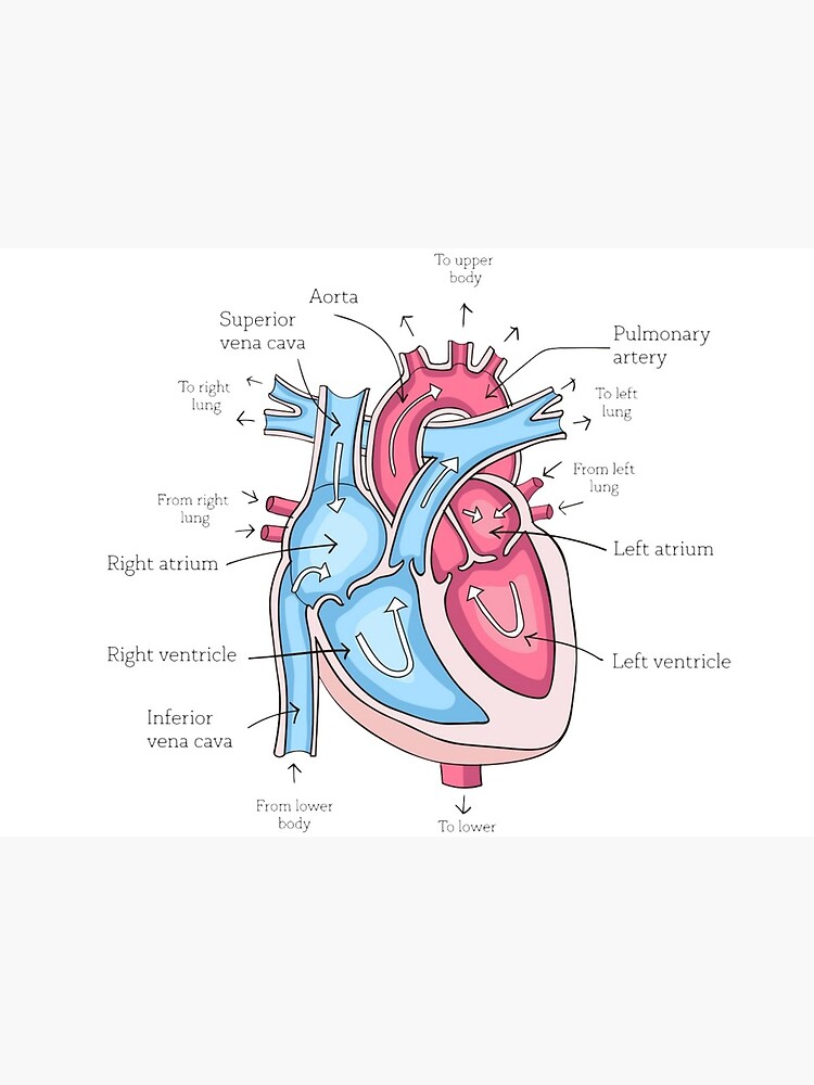
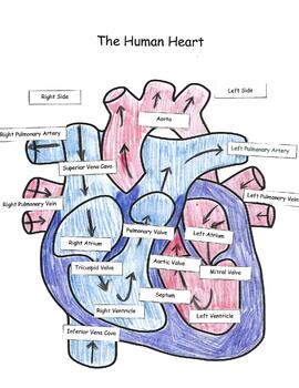
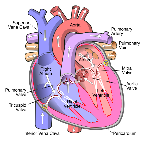
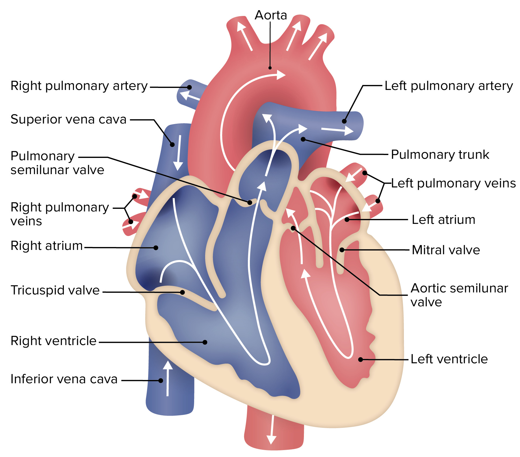
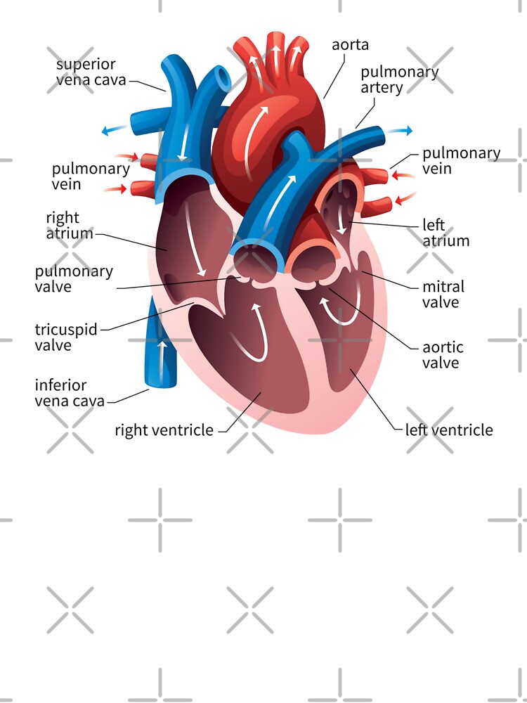
A Diagram of the Heart and Its Functioning Explained in Detail The heart blood flow diagram (flowchart) given below will help you to understand the pathway of blood through the heart.Initial five points denotes impure or deoxygenated blood and the last five points denotes pure or oxygenated blood. 1.Different Parts of the Body ↓ 2.Major Veins ↓ 3.Right Atrium ↓ 4.Right Ventricle ↓ 5.Pulmonary Artery ↓ 6.Lungs 13 parts of the human heart (and its functions) - LORECENTRAL Parts of the heart and its functions 1. Left atrium 2. Mitral Valve 3. Left Ventricle 4. Aortic sigmoid valve Right atrium 6. Tricuspid valve 7. Right ventricle 8. Pulmonary sigmoid valve 9. Atrial septal defect Interventricular partition 11. The sinus or sinoatrial node 12. Atrioventricular or Aschoff-Tawara nodule 13. Hiscules and Purkinje fibers How to Draw a Human Heart: An Easy Step-By-Step Guide - wikiHow Generally, hearts on anatomical or medical diagrams are sectioned into 2 colors: red and blue. The red represents blood going into the heart, whereas the blue represents blood leaving the heart. [8] Split the heart in half and color the left section, superior vena cava, and pulmonary artery blue, and then color the right section and aorta red. [9] heart | Structure, Function, Diagram, Anatomy, & Facts heart, organ that serves as a pump to circulate the blood. It may be a straight tube, as in spiders and annelid worms, or a somewhat more elaborate structure with one or more receiving chambers (atria) and a main pumping chamber (ventricle), as in mollusks. In fishes the heart is a folded tube, with three or four enlarged areas that correspond to the chambers in the mammalian heart. In animals ...
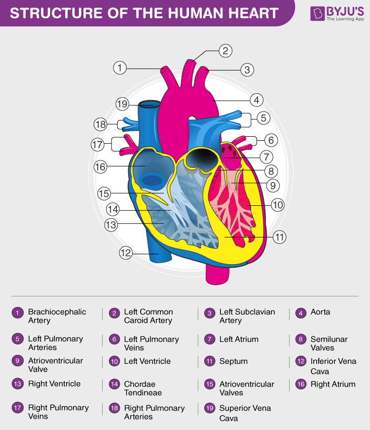
The 18 parts of the human heart, and their functions The 18 parts of the human heart and how they work 1. Myocardium 2. Endocardium 3. Pericardium 4. Right Auricle 5. Right ventricle 6. Tricuspid valve 7. Pulmonary valve 8. Left Auricle 9. Left ventricle 10. Mitral valve 11. Aortic valve 12. Tendon cords 13. Papillary muscles 14. Sinus node 15. Atrioventricular node 16. Atrioventricular fascicule 17. Human Heart - Anatomy, Functions and Facts about Heart - BYJUS The human heart is divided into four chambers, namely two ventricles and two atria. The ventricles are the chambers that pump blood and atrium are the chambers that receive the blood. Among which, the right atrium and ventricle make up the "right portion of the heart", and the left atrium and ventricle make up the "left portion of the heart." 5. Diagram of the human heart royalty-free images - Shutterstock 14,830 diagram of the human heart stock photos, vectors, and illustrations are available royalty-free. See diagram of the human heart stock video clips Image type Orientation People Artists Sort by Popular Anatomy Healthcare and Medical Icons and Graphics Diseases, Viruses, and Disorders heart medicine organ diagram hemodynamics circulatory system File : Diagram of the human heart (cropped).svg - Wikimedia Aug 08, 2022 · English: Diagram of the human heart 1. Superior vena cava 2. 4. Mitral valve 5. Aortic valve 6. Left ventricle 7. Right ventricle 8. Left atrium 9. Right atrium 10. Aorta 11. Pulmonary v
Science worksheets: Label parts of a human heart
How the Heart Works: Diagram, Anatomy, Blood Flow - MedicineNet The heart is an amazing organ. It starts beating about 22 days after conception and continuously pumps oxygenated red blood cells and nutrient-rich blood and other compounds like platelets throughout your body to sustain the life of your organs.; Its pumping power also pushes blood through organs like the lungs to remove waste products like CO2.; This fist-sized powerhouse beats (expands and ...

Heart Anatomy: Labeled Diagram, Structures, Blood Flow ...
Labelling the heart — Science Learning Hub Labelling the heart — Science Learning Hub Labelling the heart Add to collection The heart is a muscular organ that pumps blood through the blood vessels of the circulatory system. Blood transports oxygen and nutrients to the body. It is also involved in the removal of metabolic wastes. Topics Concepts Citizen science Teacher PLD Glossary Sign in

Easy way to draw human heart and label its main parts
How the Heart Works - The Heart | NHLBI, NIH - National Institutes of ... The Heart. The heart is an organ about the size of your fist that pumps blood through your body. It is made up of multiple layers of tissue. Your heart is at the center of your circulatory system. This system is a network of blood vessels, such as arteries, veins, and capillaries, that carries blood to and from all areas of your body.
Label the heart — Science Learning Hub
Dare (album) - Wikipedia Dare earned considerable income for record labels Virgin and A&M; in Virgin's case, it gave the label the first chart-topping album since Mike Oldfield's Tubular Bells in 1973. "Don't You Want Me" was the label's first ever chart-topping single. The success of Dare was responsible for saving the label from impending bankruptcy.
13+ Heart Diagram Templates – Sample, Example, Format ...
Parts Of The Human Heart | Science Trends The parts of the human heart can be broken down into four chambers, muscular walls, vessels, and a conductive system. The two upper chambers are called the atria, with lower parts called ventricles. These all work together to make up the vital function of your heart. Everybody knows that the human heart is the essential organ in our bodies.

Label the Heart Quiz
Normal chest MDCT with anatomic labels | e-Anatomy - IMAIOS Mar 10, 2022 · Pocket Atlas of Human Anatomy: 5th edition - W. Dauber, Founded by Heinz Fene Anatomical variants and notes from the author about the anatomical labeling of the thorax CT: In the lower lobe of the left lung, there is an inconstant subsuperior pulmonary segment that is seen in approximately 30% of individuals, located between the superior and ...
609,856 Human heart Images, Stock Photos & Vectors | Shutterstock
File:Diagram of the human heart (cropped).svg - Wikipedia Added shadows. Left main pulmonary artery with its first division. 07:02, 2 June 2006: 650 × 650 (26 KB) Yaddah: Diagram of the human heart, created by Wapcaplet in Sodipodi. Cropped by ~~~ to remove white space (this cropping is not the same as Wapcaplet's original crop). == See also == * Image:Diagram of the human heart.svg - original
poster of human heart anatomy with hand written labels of the main parts | Art Board Print
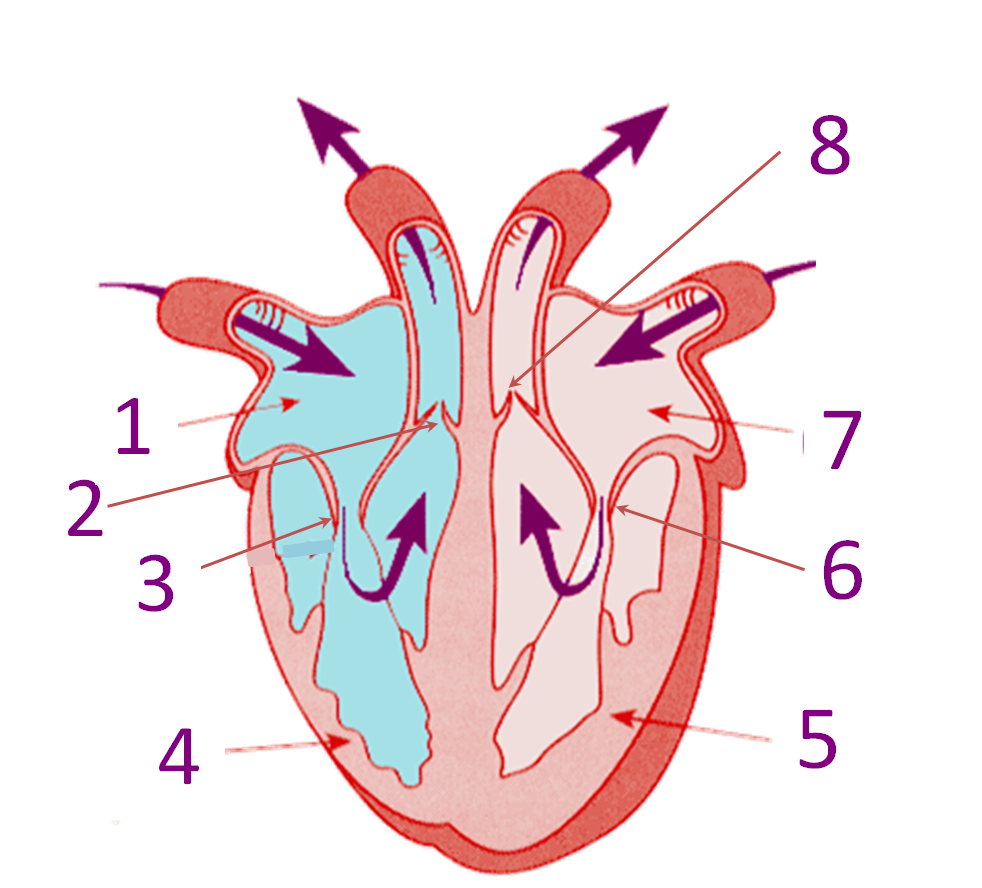
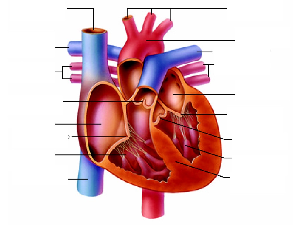
PDF Analyzing the Human Heart - Beyond the Classroom working. Its job is to pump blood to the lungs and to all of the body tissues. In this activity you will use a diagram of the heart to analyze the way in which the heart works. l. Using the following word list, label the various parts of the heart on the diagram. Right ventricle Left venfficle Upper vena cava Lower vena cava Aorta

Human Heart Labeled | Human heart diagram, Heart diagram ...
Human Heart (Anatomy): Diagram, Function, Chambers, Location in ... - WebMD The heart is a muscular organ about the size of a fist, located just behind and slightly left of the breastbone. The heart pumps blood through the network of arteries and veins called the...

Human heart with label free image download
Heart Diagram with Labels and Detailed Explanation - BYJUS Diagram of Heart. The human heart is the most crucial organ of the human body. It pumps blood from the heart to different parts of the body and back to the heart. The most common heart attack symptoms or warning signs are chest pain, breathlessness, nausea, sweating etc. The diagram of heart is beneficial for Class 10 and 12 and is frequently ...
Heart Anatomy: Labeled Diagram, Structures, Blood Flow ...
Label the heart — Science Learning Hub Label the heart Interactive Add to collection In this interactive, you can label parts of the human heart. Drag and drop the text labels onto the boxes next to the diagram. Selecting or hovering over a box will highlight each area in the diagram. pulmonary vein semilunar valve right ventricle right atrium vena cava left atrium pulmonary artery
Heart Diagram with Labels and Detailed Explanation
Heart histology: Cells and layers | Kenhub The heart is a critical organ that keeps blood moving throughout the body. Blood is an important medium that not only carries nutrients and oxygen throughout the body, but it also collects waste products and returns them to the liver and kidney for further processing and excretion.. The heart is able to achieve this autonomy based on its histological make-up.

Human Heart Circulatory System Diagram Chart Medical Educational Science Class Anatomy Corazon Veins Arteries Labels Thick Paper Sign Print Picture ...
The Anatomy of the Heart, Its Structures, and Functions - ThoughtCo The heart is the organ that helps supply blood and oxygen to all parts of the body. It is divided by a partition (or septum) into two halves. The halves are, in turn, divided into four chambers. The heart is situated within the chest cavity and surrounded by a fluid-filled sac called the pericardium. This amazing muscle produces electrical ...
Human heart cross section hi-res stock photography and images ...
Heart (Human Anatomy): Overview, Function & Structure | Biology The heart is a muscular organ that pumps blood throughout the body. It is located in the middle cavity of the chest, between the lungs. In most people, the heart is located on the left side of the chest, beneath the breastbone. The heart is composed of smooth muscle. It has four chambers which contract in a specific order, allowing the human ...
The Human Heart: Cut, Paste and Label
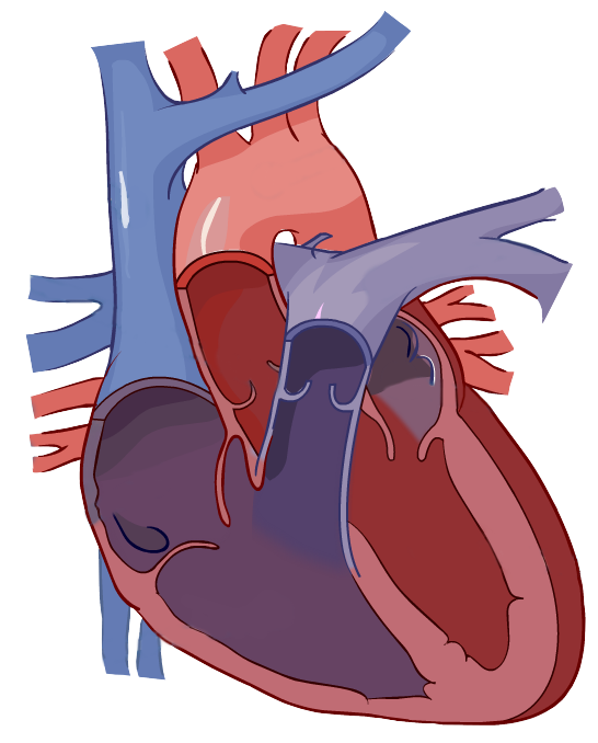
Diagram of Human Heart and Blood Circulation in It Exterior of the Human Heart A heart diagram labeled will provide plenty of information about the structure of your heart, including the wall of your heart. The wall of the heart has three different layers, such as the Myocardium, the Epicardium, and the Endocardium. Here's more about these three layers. Epicardium

External heart anatomy with labels diagram | Human heart ...
Human heart: Anatomy, function & facts | Live Science The human heart has four chambers: two upper chambers (the atria) and two lower ones (the ventricles), according to the National Institutes of Health. The right atrium and right ventricle together...

Heart Anatomy | Anatomy and Physiology II
A Labeled Diagram of the Human Heart You Really Need to See The human heart, comprises four chambers: right atrium, left atrium, right ventricle and left ventricle. The two upper chambers are called the left and the right atria, and the two lower chambers are known as the left and the right ventricles. The two atria and ventricles are separated from each other by a muscle wall called 'septum'.

Human Heart Circulatory System Diagram Chart Medical Educational Science Class Anatomy Corazon Veins Arteries Labels Black Wood Framed Art Poster ...
Heart Pictures, Diagram & Anatomy | Body Maps - Healthline The average human heart weighs between 6 and 11 ounces. The muscle is strong enough to pump up to 2,000 gallons — as much as a fire department's tanker truck — of blood through one's body ...

Human Heart Anatomy Infographic Diagram Stock Vector ...
Human Genome Project Results Nov 12, 2018 · By using mRNA as a template, scientists use enzymatic reactions to convert its information back into cDNA and then clone it, creating a collection of cDNAs, or a cDNA library. These libraries are important to scientists because they consist of clones of all protein-encoding DNA, or all of the genes, in the human genome.
Heart Anatomy: Labeled Diagram, Structures, Blood Flow ...
Prepare a drawing of the human heart and label its parts. | Quizlet Draw a diagram of the heart showing the three layers composing its wall and its four chambers. Label each. Show where the AV and semilunar valves are. Show and label all blood vessels entering and leaving the heart chambers.
File:Diagram of the human heart (cropped).svg - Wikipedia
Parts Of The Heart - ProProfs Quiz

Human Heart Diagram Labeled | Science Trends
Heart: Anatomy | Concise Medical Knowledge
Draw the structure of a human heart and label its parts ...

Human Heart Anatomy High-Res Vector Graphic - Getty Images

File:Heart diagram-en.svg - Wikimedia Commons
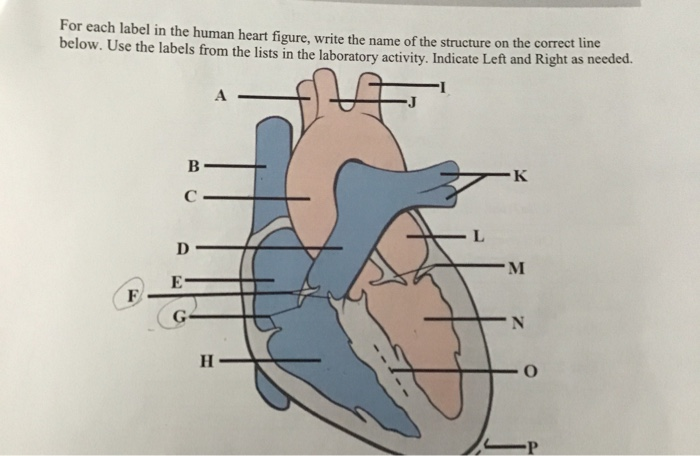
Solved for each label in the human heart figure,write the ...

Activity
Label the Human Heart | eCampusOntario H5P Studio
Heart: Anatomy and Function

Given Alongside is a Diagram of Human Heart Showing Its ...

YR 8 Topic 2 Circulatory System - AMAZING WORLD OF SCIENCE ...

Given alongside is a diagram of the human heart showing its internal structure. Label the parts ...

Heart Anatomy: Labeled Diagram, Structures, Blood Flow ...

Draw a diagram of the human heart and label its parts.

Heart Anatomy | Anatomy and Physiology II
poster of human heart anatomy with hand written labels of the main parts | Kids T-Shirt
Free Heart Diagram Unlabeled, Download Free Heart Diagram ...
6f3bdbd66d563734dd2473aeb23411 ...
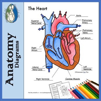
Heart Diagrams for Labeling and Coloring, With Reference ...

Human Body - The Human Heart
Post a Comment for "42 the human heart and its labels"