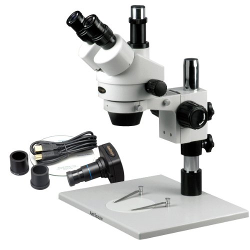
41 dissecting microscope diagram with labels
rsscience.com › stereo-microscopeParts of Stereo Microscope (Dissecting microscope) – labeled ... If you would like to learn optical components of a compound microscope, please visit Compound Microscope Parts – Labeled Diagram and their Functions, and this article. How to use a stereo (dissecting) microscope. Follow these steps to put your stereo microscopes in work: 1. Set your microscope on a tabletop or other flat sturdy surface where ... › articles › s41586/022/05028-xA physical wiring diagram for the human immune system | Nature Aug 03, 2022 · For imaging, a PerkinElmer Opera Phenix automated spinning-disk confocal microscope was used and each well of a 348-well plate was imaged at 20× magnification with 5 × 5 non-overlapping images ...
Dissecting the treatment-naive ecosystem of human melanoma … Web07.07.2022 · Introduction. Melanoma brain metastases (MBMs) are the third most common cause of brain metastases after carcinomas of the lung and breast (Eichler et al., 2011) and lead to significant morbidity and mortality (Davies et al., 2011).While treatment with combination immune checkpoint blockade can be effective in patients with MBM (Tawbi …

Dissecting microscope diagram with labels
› science › articleDissecting the treatment-naive ecosystem of human melanoma ... Jul 07, 2022 · Animals were anesthetized with a ketamine (100 mg/kg) and xylazine (10 mg/kg) cocktail. Organs of interest were dissected and placed in a plate containing HBSS (Hank's buffered salt solution) on ice. GFP+ metastases were visualized using a Leica M205 FA fluorescence stereo (dissecting) scope. GFP+ areas were dissected away from the organ ... Single-cell transcriptome atlas reveals ... - Wiley Online Library Web10.07.2022 · To determine the thickness and degree of lignification of leaf cell, the untreated slices were also observed under the confocal laser scanning microscope (Lecia DMi8, Germany). The excitation light wavelength of the blue, green and red channel was 405 nm, 488 nm and 488 nm, respectively and the emission light wavelength range was 430–480 … Heterogeneity in endothelial cells and widespread venous ... - Nature Web25.01.2022 · Images in red boxes are shown at high magnification. The diagram on the upper left indicates the position of sections. nt, neural tube; ACV, anterior cardinal vein; DA, dorsal aorta. Scale bars ...
Dissecting microscope diagram with labels. Latest Breaking News, Headlines & Updates | National Post WebRead latest breaking news, updates, and headlines. Get information on latest national and international events & more. onlinelibrary.wiley.com › doi › 10Single‐cell transcriptome atlas reveals developmental ... Jul 10, 2022 · To determine the thickness and degree of lignification of leaf cell, the untreated slices were also observed under the confocal laser scanning microscope (Lecia DMi8, Germany). The excitation light wavelength of the blue, green and red channel was 405 nm, 488 nm and 488 nm, respectively and the emission light wavelength range was 430–480 nm ... › tech-article › refractometersWhat is a Refractometer & How Does it Work - Cole-Parmer Jul 12, 2022 · Measurements are read at the point where the prism and solution meet. With a low concentration solution, the refractive index of the prism is much greater than that of the sample, creating a large refraction angle and a low reading ("A" on diagram). The reverse would happen with a high concentration solution ("B" on diagram). COVID-19 immune features revealed by a large-scale single-cell ... Web01.04.2021 · Single-cell RNA sequencing (scRNA-seq) is powerful at dissecting the immune responses and has been applied to COVID-19 studies (Cao et al., 2020; Chua et al., 2020; Fan et al., 2020; Su et al., 2020; Wen et al., 2020; Xie et al., 2020; Zhang et al., 2020a, 2020b). While the current single-cell studies of COVID-19 have provided …
COVID-19 immune features revealed by a large-scale single-cell ... Web03.02.2021 · IGHV genes differentially used by moderate or severe COVID-19 patients compared with healthy controls and their intersections are shown with different colors. Venn diagram is used to show their overlaps with those published SARS-CoV-2 antibodies. Adjusted p values < 0.05 are indicated (two-sided unpaired Wilcoxon test). Parts of Stereo Microscope (Dissecting microscope) – labeled diagram ... WebCompared to a compound microscope where the objectives attached to the nosepiece can be seen and identified individually (based on color bands and their respective labels), the objectives of a dissecting microscope are located in a cylindrical cone and, therefore, are not directly seen. For the stereo microscope that comes with multiple objective lens sets … (PDF) Principles and Techiniques of Biochemistry and Molecular … WebPrinciples and Techiniques of Biochemistry and Molecular Biology 7th ed wilson walker A physical wiring diagram for the human immune system | Nature Web03.08.2022 · The human immune system is composed of a distributed network of cells circulating throughout the body, which must dynamically form physical associations and communicate using interactions between ...
› science › articleCOVID-19 immune features revealed by a large-scale single ... IGHV genes differentially used by moderate or severe COVID-19 patients compared with healthy controls and their intersections are shown with different colors. Venn diagram is used to show their overlaps with those published SARS-CoV-2 antibodies. Adjusted p values < 0.05 are indicated (two-sided unpaired Wilcoxon test). What is a Refractometer & How Does it Work - Cole-Parmer Web12.07.2022 · Refractometer FAQs What is a Refractometer? A refractometer is a simple instrument used for measuring concentrations of aqueous solutions such as gases, liquids, and translucent solids. Different types of refractometers are available depending on the application. Refractometers can be handheld, compact, benchtop, Abbe, and Brix as well … › 33654457 › Principles_and_TechiniPrinciples and Techiniques of Biochemistry and Molecular ... Enter the email address you signed up with and we'll email you a reset link. Heterogeneity in endothelial cells and widespread venous ... - Nature Web25.01.2022 · Images in red boxes are shown at high magnification. The diagram on the upper left indicates the position of sections. nt, neural tube; ACV, anterior cardinal vein; DA, dorsal aorta. Scale bars ...
Dissecting Microscopes - MicroscopeGenius.com
Single-cell transcriptome atlas reveals ... - Wiley Online Library Web10.07.2022 · To determine the thickness and degree of lignification of leaf cell, the untreated slices were also observed under the confocal laser scanning microscope (Lecia DMi8, Germany). The excitation light wavelength of the blue, green and red channel was 405 nm, 488 nm and 488 nm, respectively and the emission light wavelength range was 430–480 …
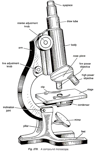
Compound Microscope Drawing With Label - Micropedia
› science › articleDissecting the treatment-naive ecosystem of human melanoma ... Jul 07, 2022 · Animals were anesthetized with a ketamine (100 mg/kg) and xylazine (10 mg/kg) cocktail. Organs of interest were dissected and placed in a plate containing HBSS (Hank's buffered salt solution) on ice. GFP+ metastases were visualized using a Leica M205 FA fluorescence stereo (dissecting) scope. GFP+ areas were dissected away from the organ ...

30 Label Parts Of Microscope - Labels Database 2020
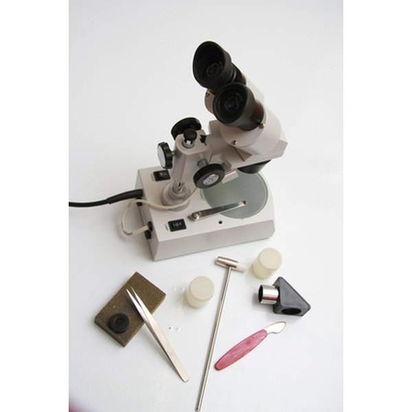
Parts of the Dissecting Microscope | Synonym
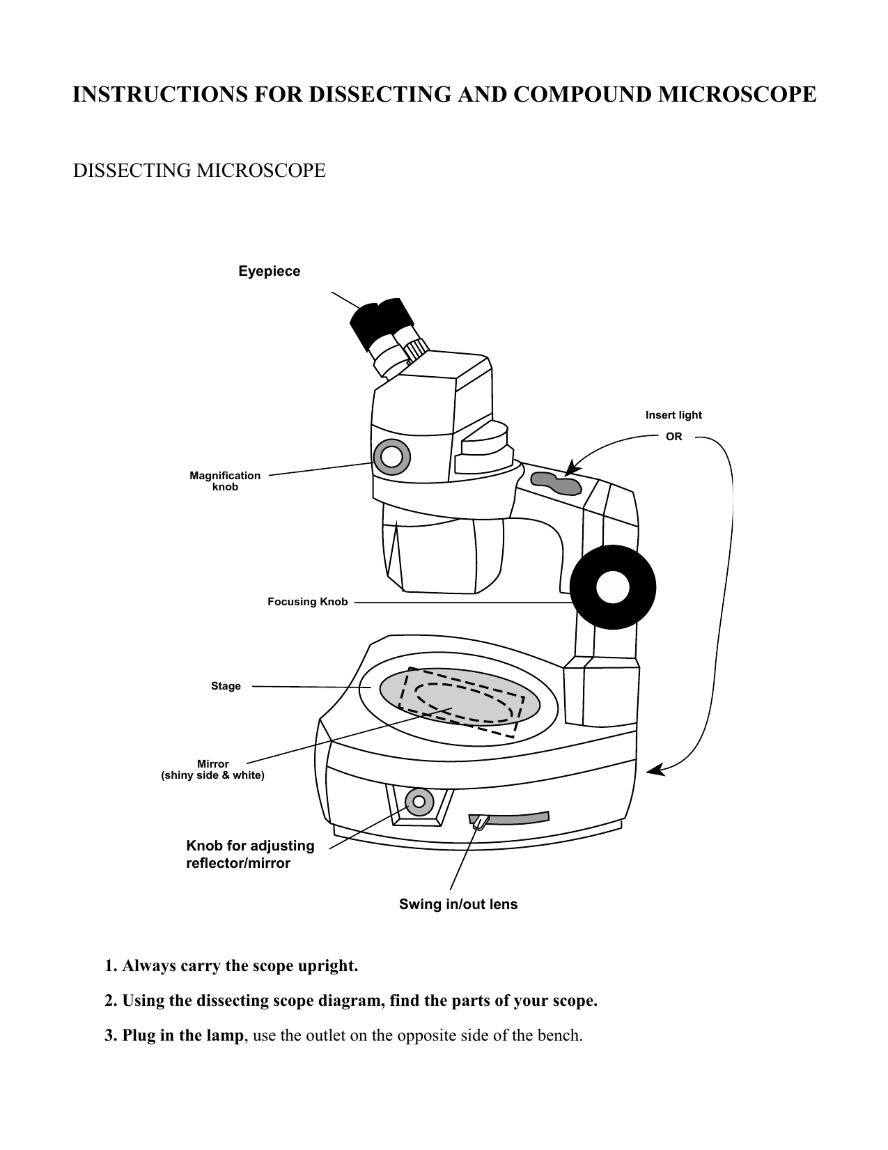
Drawing Simple Dissecting Microscope Diagram - Micropedia
Hetal & Craig - Armstrong Plant Biology Lab
Difference Between Compound & Dissecting Microscopes | Sciencing
![How to Use a Microscope: Lesson for Kids - Science Class [2021] | Study.com](https://study.com/cimages/multimages/16/labeledmicroscopeimage.jpg)
How to Use a Microscope: Lesson for Kids - Science Class [2021] | Study.com

Microscope World Blog: November 2013
Homeschool Supplies & Science Supply List - Homeschool Den
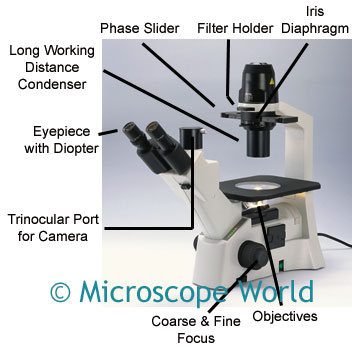
Microscope World Blog: Inverted Biological Microscopes

Dissecting Microscope With Labeled Parts - Micropedia

Compound Microscope Unlabeled - Micropedia

All Saints Online
Parts of the Dissecting Microscope | Synonym

microscope labeled diagram simple microscope beauteous labeled - Top Label Maker

Chapter 1 Questions PPT - BIOLOGY JUNCTION
Post a Comment for "41 dissecting microscope diagram with labels"