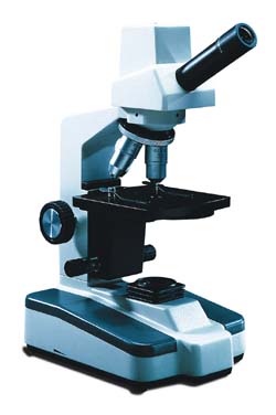
40 light microscope with labels
Labelled Diagram Of A Light Microscope - GlobalSpec Products/Services for Labelled Diagram Of A Light Microscope Microscopes - (706 companies) ...and transmission electron microscopes. Acoustic and ultrasonic microscopes use sound waves to create images of the sample. Compound microscopes use a single light path. These types of microscopes can have a single eyepiece (monocular) or a dual eyepiece... Labeling the Parts of the Microscope Labeling the Parts of the Microscope This activity has been designed for use in homes and schools. Each microscope layout (both blank and the version with answers) are available as PDF downloads. You can view a more in-depth review of each part of the microscope here. Download the Label the Parts of the Microscope PDF printable version here.
Compound Microscope Parts, Functions, and Labeled Diagram Compound Microscope Definitions for Labels. Eyepiece (ocular lens) with or without Pointer: The part that is looked through at the top of the compound microscope. Eyepieces typically have a magnification between 5x & 30x. Monocular or Binocular Head: Structural support that holds & connects the eyepieces to the objective lenses.

Light microscope with labels
Microscope Types (with labeled diagrams) and Functions This is an advanced microscope that has specific application in viewing, observing and measuring the optical thickness and phase of completely transparent specimens and objects. A tiny interferometer is used and a specimen is placed on beam path of it. This path is split and then rejoined to create two superimposed images of the specimen in focus. Light microscopes - Cell structure - Edexcel - BBC Bitesize Microscopes are used to produce magnified images. There are two main types of microscope: light microscopes are used to study living cells and for regular use when relatively low magnification and... A quick guide to light microscopy in cell biology - PMC This allows the labels to be targeted to the molecule(s) of interest, either by genetically encoding a fluorescent protein or by binding a fluorescently labeled antibody. ... Finally, should you have access to a light sheet microscope or other specialized microscope, learn its capabilities and consider whether it is a good fit for your ...
Light microscope with labels. › microscopy › intSmart Microscope for Lab Routine and Research - ZEISS Acquiring fluorescent images has never been so easy. Combine Axioscope 5 with the LED light source Colibri 3 and the sensitive, standalone microscope camera Axiocam 202 mono to have the perfect setup for easy multichannel fluorescence documentation. Switch effortlessly between the channels for UV, blue, green and red excitation. Light Microscope: Definition, Uses & Parts - Study.com A light microscope uses focused light and lenses to magnify a specimen, usually a cell. In this way, a light microscope is much like a telescope, except that instead of the object being very large ... Student's Guide: How to Use a Light Microscope Considered one of the most versatile techniques of optical imaging, fluorescence microscopy uses a fluorescent substance (e.g., fluorochromes or fluorophores) to tag or label a specimen of interest. A fluorescent microscope uses a high-intensity illuminator which then excites the fluorophores in the sites of interest. Compound Microscope Parts - Labeled Diagram and their Functions - Rs ... The eyepiece (or ocular lens) is the lens part at the top of a microscope that the viewer looks through. The standard eyepiece has a magnification of 10x. You may exchange with an optional eyepiece ranging from 5x - 30x. [In this figure] The structure inside an eyepiece. The current design of the eyepiece is no longer a single convex lens.
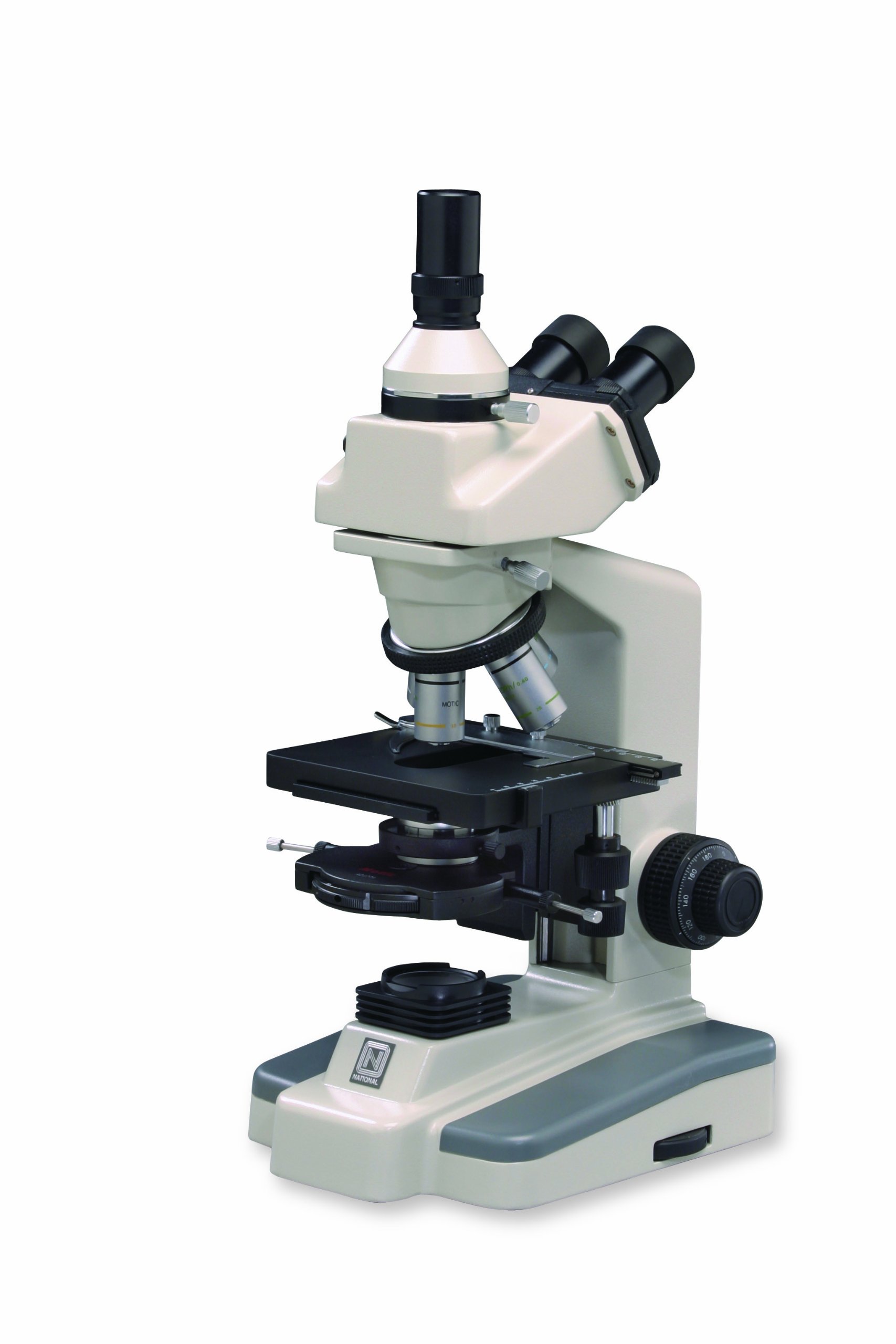
An Introduction to the Light Microscope, Light Microscopy Techniques ... a) standard upright microscope indicating (1) e yepiece (ocular lens), (2) objective turret, revolver, or revolving nose piece (to hold multiple objective lenses), (3) objective lenses, focus knobs (to move the stage) (4) coarse adjustment, (5) fine adjustment, (6) stage (to hold the specimen), (7) light source (a light or a mirror), (8) … Microscope labels Flashcards | Quizlet Microscope label Learn with flashcards, games, and more — for free. › AmScope-LED-144W-ZK-AdjustableAmScope LED-144W-ZK White Adjustable 144 LED Ring Light ... Buy AmScope LED-144W-ZK White Adjustable 144 LED Ring Light Illuminator for Stereo Microscope ... DYMO Authentic LW Large Multi-Purpose Labels for LabelWriter Label ... Microscope Labeling - The Biology Corner 1) Start with scanning (the shortest objective) and only use the COARSE knob . Once it is focused… 2) Switch to low power (medium) and only use the COARSE knob . You may need to recenter your slide. Once it is focused.. 3) Switch to high power (long objective).
Label the Light Microscope - Labelled diagram Eyepiece, Light Source, Base, Stage, Stage Clips, Fine Focus, Coarse Focus, Arm, Objective Lens. ... Label the Light Microscope. Share Share by Nquinn805. Like. Edit Content. Embed. More. Leaderboard. Show more Show less . This leaderboard is currently private. Click Share to make it public. This leaderboard has been disabled by the resource ... en.wikipedia.org › wiki › Electron_microscopeElectron microscope - Wikipedia An electron microscope is a microscope that uses a beam of accelerated electrons as a source of illumination. As the wavelength of an electron can be up to 100,000 times shorter than that of visible light photons, electron microscopes have a higher resolving power than light microscopes and can reveal the structure of smaller objects. Light Microscope- Definition, Principle, Types, Parts, Labeled Diagram ... A light microscope is a biology laboratory instrument or tool, that uses visible light to detect and magnify very small objects and enlarge them. They use lenses to focus light on the specimen, magnifying it thus producing an image. The specimen is normally placed close to the microscopic lens. Compound Light Microscopes | Products | Leica Microsystems Light microscope solutions help suppliers and device manufacturers achieve fast and precise inspection and analysis for semiconductor wafer processing. Conformity to the defined specifications during semiconductor device manufacturing is critical for reliability. ... (CRS) is a powerful approach for label-free, chemically specific imaging. It ...
33 Label A Compound Light Microscope - Labels Database 2020
Light Microscope: Functions, Parts and How to Use It To use a light microscope, you can follow the steps below carefully. Start with a low lens and a clean slide. The microscope stage should be lowered as low as possible. Center the slide so that the specimen is under the objective lens. Use the coarse adjustment knob to get a general focus. Then slowly move up the stage until focus is achieved.
Bio F4 Cell Organel
Label a Microscope - Storyboard That In this activity, students will create a poster of a microscope with labeled parts. Students will identify and describe the microscope parts and functions. This is an awesome activity to complete at the beginning of either the school year or the unit on basic cells. ... Provides light to illuminate the specimen, sometimes a mirror is also used ...
33 Label All Indicated Parts Of The Microscope - Labels Database 2020
Light Microscope - an overview | ScienceDirect Topics The polarized light microscope is a highly specialized tool quite unlike a medical or biological microscope. Their only similarity is in presenting a magnified image of the sample to the user. The basic element in any microscope is the stand. The PLM stand should be quite sturdy and be capable of accepting a variety of auxiliary components.
Labeling A Compound Light Microscope - ClipArt Best
Microscope Parts and Functions Microscope Parts and Functions With Labeled Diagram and Functions How does a Compound Microscope Work?. Before exploring microscope parts and functions, you should probably understand that the compound light microscope is more complicated than just a microscope with more than one lens.. First, the purpose of a microscope is to magnify a small object or to magnify the fine details of a larger ...
Quia - Protist Vocabulary
Microscope Labeling - The Biology Corner Students label the parts of the microscope in this photo of a basic laboratory light microscope. Can be used for practice or as a quiz. Name_____ Microscope Labeling . Microscope Use: 15. When focusing a specimen, you should always start with the _____ objective.

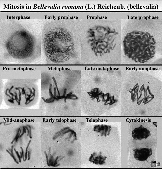
Histology Drawings: January 2014
› books › NBK26880Looking at the Structure of Cells in the Microscope ... Many light-microscope techniques are available for observing cells. Cells that have been fixed and stained can be studied in a conventional light microscope, while antibodies coupled to fluorescent dyes can be used to locate specific molecules in cells in a fluorescence microscope. Living cells can be seen with phase-contrast, differential ...

Label Compound Light Microscope - ClipArt Best
Label the microscope — Science Learning Hub All microscopes share features in common. In this interactive, you can label the different parts of a microscope. Use this with the Microscope parts activity to help students identify and label the main parts of a microscope and then describe their functions. Drag and drop the text labels onto the microscope diagram.
compound light microscope clipart 10 free Cliparts | Download images on Clipground 2021
Label the light microscope | Teaching Resources Label the light microscope. Subject: Biology. Age range: 11-14. Resource type: Worksheet/Activity (no rating) 0 reviews. Science Resources. 4 1 reviews. ... Low ability worksheet, label the main parts of a microscope, key terms given at the bottom of the worksheet. Creative Commons "Attribution"

Microscope Picture To Label - Micropedia
› products › microscopeMicroscope Objective Lens | Products | Leica Microsystems The objective lens is a critical part of the microscope optics. The microscope objective is positioned near the sample, specimen, or object being observed. It has a very important role in imaging, as it forms the first magnified image of the sample. The numerical aperture (NA) of the objective indicates its ability to gather light and largely determines the microscope’s resolution, the ...

Rens blog : Science, cells
Label the Microscope | Microscope parts, Teaching biology, Biology labs Label the Microscope. Description Worksheet identifying the parts of the compound light microscope. Answer key: 1. Body tube 2. Revolving nosepiece 3. Low power objective 4. Medium power objective 5. High power objective 6.
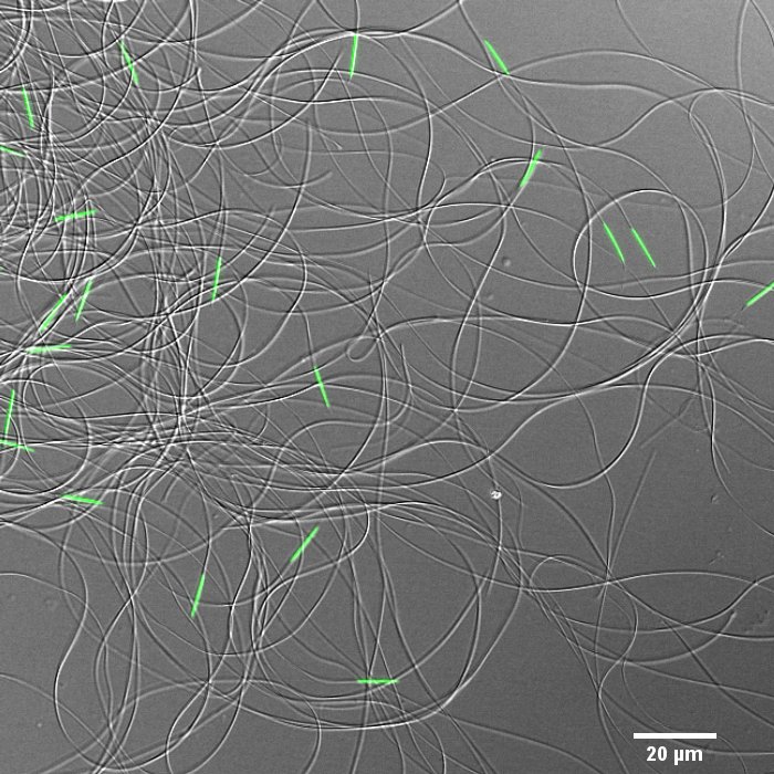
Drosophila sperm cells | Advanced Light Microscopy Facility
Simple Microscope - Diagram (Parts labelled), Principle, Formula and Uses A simple microscope consists of Optical parts Mechanical parts Labeled Diagram of simple microscope parts Optical parts The optical parts of a simple microscope include Lens Mirror Eyepiece Lens A simple microscope uses biconvex lens to magnify the image of a specimen under focus.
Search in gallery
rsscience.com › stereo-microscopeParts of Stereo Microscope (Dissecting microscope) – labeled ... A stereo microscope allows you to see the surface of specimens with a 3-dimensional view. Under a stereo microscope, you can see the metallic texture and colors of the mosquito’s compound eyes. In contrast, the light has to pass through the specimen to form the image under a compound microscope.
Labeling A Compound Light Microscope - ClipArt Best
Parts of the Microscope with Labeling (also Free Printouts) Parts of the Microscope with Labeling (also Free Printouts) A microscope is one of the invaluable tools in the laboratory setting. It is used to observe things that cannot be seen by the naked eye. Table of Contents 1. Eyepiece 2. Body tube/Head 3. Turret/Nose piece 4. Objective lenses 5. Knobs (fine and coarse) 6. Stage and stage clips 7. Aperture

32 Compound Light Microscope With Label - Labels For You
Light Microscope - an overview | ScienceDirect Topics The light microscope is an instrument for visualizing fine detail of an object. It does this by creating a magnified image through the use of a series of glass lenses, which first focus a beam of light onto or through an object, and convex objective lenses to enlarge the image formed.

microscopic organisms - nwnature.net
Parts of a microscope with functions and labeled diagram Microscopic illuminator - This is the microscopes light source, located at the base. It is used instead of a mirror. It captures light from an external source of a low voltage of about 100v. Condenser - These are lenses that are used to collect and focus light from the illuminator into the specimen.

Compound Light Microscope Labeled - Made By Creative Label
www2.nau.edu › lrm22 › lessonsMicroscope Notes - Northern Arizona University Microscope Drawings. When drawing what you see under the microscope, follow the format shown below. It is important to include a figure label and a subject title above the image. The species name (and common name if there is one) and the magnification at which you were viewing the object should be written below the image.
Post a Comment for "40 light microscope with labels"